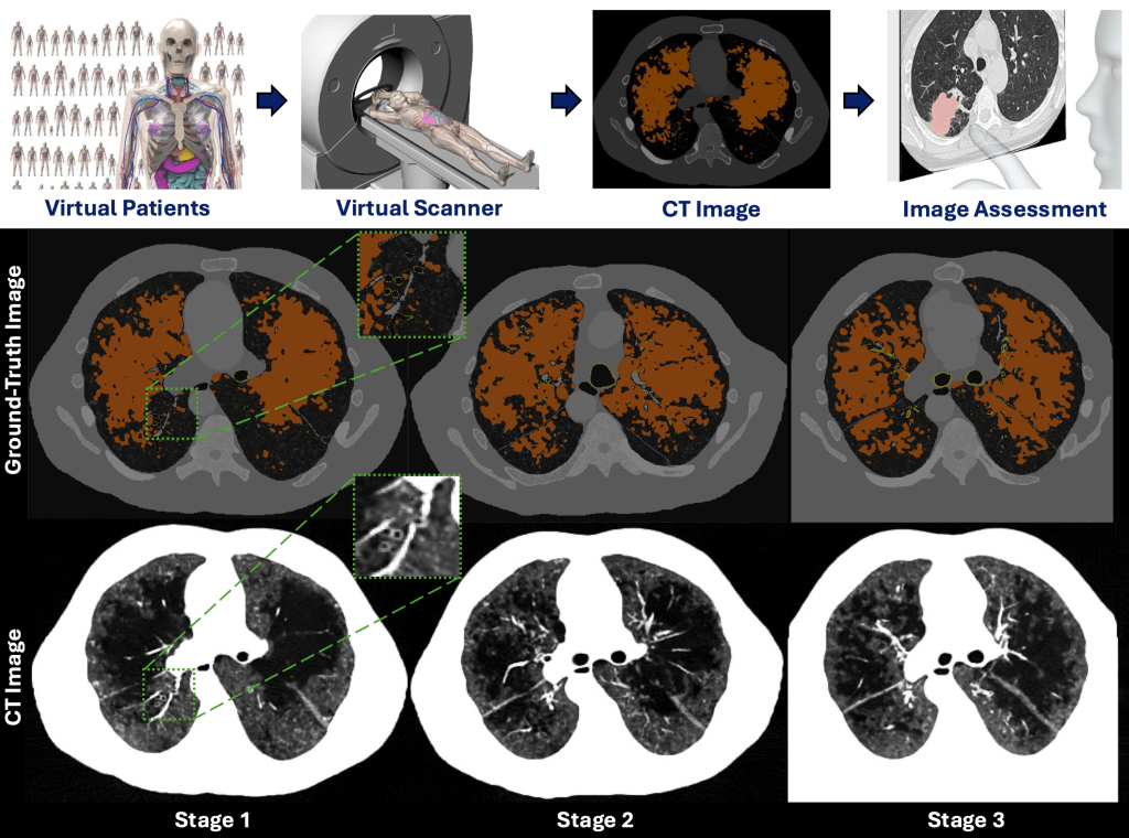
Using virtual/in-silico imaging trials with digital twins to assess the quantitative performance of CT scanners and acquisition protocols for COPD (emphysema and bronchitis) biomarkers, ensuring accuracy and consistency in both cross-sectional and longitudinal evaluations.
Study 1: Quantitative accuracy of CT protocols for cross-sectional and longitudinal assessment of COPD
Citation:
Mridul Bhattarai, Daniel W. Shin, Fong Chi Ho, Saman Sotoudeh-Paima, Ilmar Hein, Steven Ross, Naruomi Akino, Kirsten L. Boedeker, Ehsan Samei, Ehsan Abadi, “Quantitative accuracy of CT protocols for cross-sectional and longitudinal assessment of COPD: a virtual imaging study,” Proc. SPIE 13405, Medical Imaging 2025: Physics of Medical Imaging, 134054B (8 April 2025); https://doi.org/10.1117/12.3046945
Study 2: Edge-on irradiated silicon-based photon-counting CT vs. energy-integrating CT for bronchitis quantification
Innovation/Impact
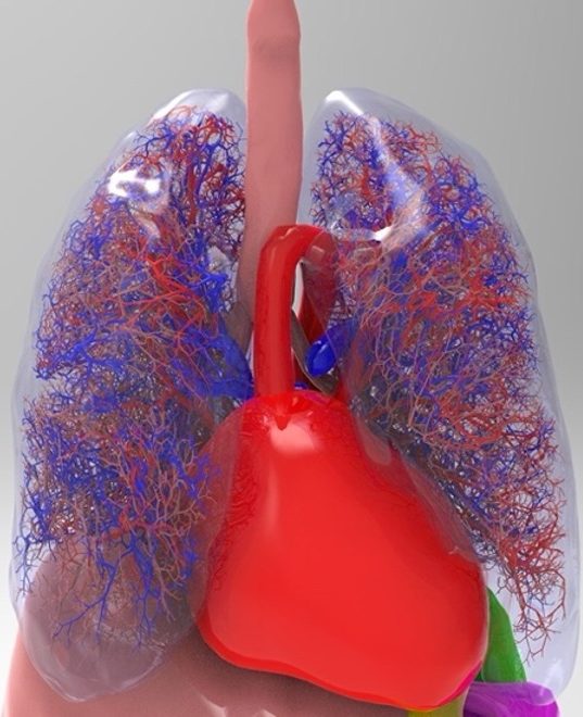
Chronic obstructive pulmonary disease (COPD) is a major cause of death in the US, characterized by emphysema and bronchitis. Bronchitis is the inflammation of the bronchial tubes (airways), and airway measurements using quantitative CT can provide an objective assessment of bronchitis severity. However, the accuracy of this measurement is limited by the spatial resolution and image noise of the CT imaging systems. The emerging photon-counting CT (PCCT) technology has the potential to improve airway quantification due to its superior noise and spatial resolution performance. To quantify its benefits, it is important to do a systematic task-specific evaluation of PCCT against energy-integrating CT (EICT) for airway measurements. However, conducting such studies on real patients is not cost-effective and has ethical limitations as well. Additionally, acquiring ground-truth information from patient images is also not feasible, which is required to measure the degradation caused by the scanner or the imaging parameters. The virtual imaging trial framework can simulate real CT scanning of anthropomorphic-computational human phantoms (XCAT) with varying severity of bronchitis. This framework was used in this study to compare the quantitative performance of deep silicon-based PCCT with conventional EICT for airway measurements and identify the optimal CT imaging parameters for accurate bronchitis quantification.
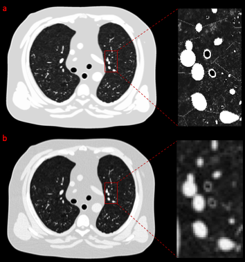
(b) CT image acquired at 350 mA and reconstructed using weighted filtered back projection and standard kernel.
[W/L] = [1000/-450].
